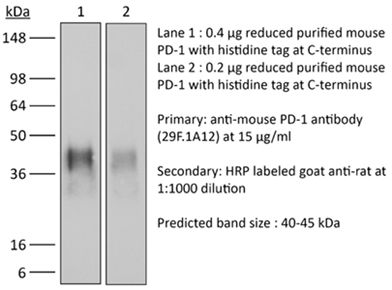
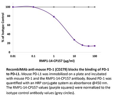
The29F.1A12™monoclonal antibody reacts with mouse PD-1(programmed death-1)also known as CD279。PD-1is a50-55kDa cell surface receptor encoded by the Pdcdcd1gene that belongs to the CD28family of the Ig superfamily.PD-1is transiently expressed on CD4and CD8thymocytes as well as activated T and velymphocytes and myeloid cells.PD-1expression declines after successfuinal in in in in in in of Addigations cd1mRNA is expressed in developing B lymphocytes during the pro-B-cell stage.PD-1’s structure includes a ITIM(immunoreceptor tyrosine-based inhibitory motif)suggesting that PD-1negatively regulates TCR signals。PD-1signals via binding its two ligands,PD-L1and PD-L2both members of the B7family.Upon ligand binding,PD-1signaling inhibits T-cell activation,leading to reduced proliferation,cytokine production,and T-cell death.Additionally,PD-1is know to play proliferation,cytokine production production,T-cel death.Additionaly to plance of opertivance immune disease in mice as PD-1knockout animals show dilated cardiomyopathy,splenomegaly,and loss of peripheral tolerance.Induced PD-L1expression is common in many tumors including squamous cell carcinoma,colon adenocarcinoma,and breast adenocarcinoma.PD-L1overexpression ression ression ress increased resstance of tumoted is.In mouse models of melanoma,tumor growth can be transiently arrested via treatment with antibodies which block the interaction between PD-L1and its receptor PD-1.For these reasons anti-PD-1mediated immunotherapies are currently being explored as cancer treatments.Like the RMP1-14 and J43 antibodies the F.12 bloown PD-1 to its ligandsin vivo。
The antibody solution should be stored at the stock concentration at4°C。Do not freeze。
*其他quality control measures for ourInVivoPlus™products include advanced binding validation,murine pathogen screening,protein aggregation screening,and ultra-low endotoxin levels.The superior quality of ourInVivoPlus™products will meet and exceed the strict demands and rigorous standards required forin vivoresearch.Learn more about theInVivoPlus™differencehere,here。
Pancreatic ductal adenocarcinoma(PDA)is characterized by immune tolerance and immunotherapeutic resistance。We discovered upregulation of receptor-interacting serine/threonine protein kinase1(RIP1)in tumor-associated macrophages(TAMs)in PDA。To study its role in oncogenic progression,we developed a selective small-molecule RIP1 inhibitor with highin vivoexposure.Targeting RIP1reprogrammed TAMs toward an MHCII(hi)TNFAlpha(+)IFNgamma(+)immunogenic phenotype in a STAT1-dependent manner。RIP1 inhibition in TAMs resulted in cytotoxic T cell activation and Thelper cell differentation toward a mixed Th1/Th17 phenotype,leading to tumor immunity in mice and in organotypic models of human PDA.Targing RIP1synergized with PD1-and inducible co-stimulator-based immunotheries.Targepopties预应力构件co-association with RIP3.Collectively,our work describes RIP1as a checkpoint kinase governing tumor immunity。
in vivoPD-1/PD-L信号
Gordon,S.R.,et al.(2017)。“PD-1expression by tumour-associated macrophages inhibits phagocytosis and tumour immunity”Nature545(7655):495-499。PubMed
Programmed cell death protein1(PD-1)is an immune checkpoint receptor that is upregulated on activated T cells for the induction of immune tolerance。Tumour cells frequently overexpress the ligand for PD-1,programmed cell death ligand1(PD-L1),facilitating their escape from the immune system.Monoclonal antibodies that block the interaction between PD-1and PD-L1,by binding to either the ligand or receptor,have shown notable clinical efficy in patients with a variety of cancers,including melanoma,colorectal cancer,non-small-Holking’s mphoma.Although it iswell established that PD-1-PD-L1blockade activates T cells,little is known about the role that this pathway may have in tumour-associated macrophages(TAMs)。Here we show that both mouse and human TAMs express PD-1.TAM PD-1expression increases over time in mouse models of cancer and with increasing disease stage in primary human cancers.TAM PD-1expression correlates negatively with phagocytic potency against tumour cells,and blockadeof-PD1in vivoincreases macrophage phagocytosis,reduces tumour growth and lengthens the survival of mice in mouse models of cancer in a macrophage-dependent fashion.This suggests that PD-1-PD-L1 therapies may also function through adirect effect on macrophages,with substantial implications for the withs。
in vivoPD-1/PD-L signaling,Flow Cytometry
Koyama,S.,et al.(2016)。“Adaptive resistance to therapeutic PD-1blockade is associated with upregulation of alternative immune checkpoints”Nat Commun7:10501。PubMed
PD-1:PD-L1 immune checkpoint in lung cancer,resistance to these therapies has increasingly been observed.In this study,to elucidate mechanisms of adaptive resistance,we analyse the tumour immune microenvironment in the context of anti-PD-1therapy in two fully immunocompetent mouse of lung adenocarcinoma.Intumours progressing follong-Panty we observe upregulation of alternative immune checkpoints,notably T-cell immunoglobulin mucin-3(TIM-3),in PD-1antibody bound T cells and demonstrate a survival advantage with addition of a TIM-3blocking antibody following failure of PD-1blockade.Two patients who developed adaptive resistance to anti-PD-1treatment also show a similar TIM-3upregulation in blockantibody-treatment分度式通风装置of TIM-3and other immune checkpoints may be targetable biomarkers associated with adaptive resistance to PD-1blockade。
in vivoPD-1/PD-L信号
Koyama,S.,et al.(2016)。“STK11/LKB1Deficiency Promotes Neutrophil Recruitment and Proinflammatory Cytokine Production to Suppress T-cell Activity in the Lung Tumor Microenvironment”Cancer Res76(5):999-1008。PubMed
STK11/LKB1is among the most commonly inactivated tumor suppressors in non-small cell lung cancer(NSCLC),especially in tumors harboring KRAS mutations.Many oncogenes promote immune escape,undermining the effectiveness of immunotherapies,but it is unclear whether the inactivation of tumor suppressor genes,such as STK11/LKB1,exerts similar effects.In this study,we investigated the consequences of KBs croenvironment ina mouse model of KRAS-driven NSCLC.Genetic ablation of STK11/LKB1 resulted in accumulation of neutrophils with T-cell-suppressive effects,along with a corresponding increase in the expression of T-cell exhaustion markers and tumor-promoting cytokines.The number of tumor-infiltratialdumps in LKB1-deficent mouse and humantumors.Furthermore,STK11/LKB1-inactivating mutations were associated with reduced expression of PD-1ligand PD-L1in mouse and patient tumors as well as in tumor-derived cell lines.Consist with these results,PD-1-targeting antibodies were ineffective against Lkb1-deficiturs,Interas-deficent mice with an IL6-neutralizing antibody or aneutrophil-depleting antibody yielded therapeutic benefits associated with reduced neutrophil accumulation and proinflammatory cytokine expression.Our findings illustrate how tumor suppressor mutations can modulate the immune milieu of the tumor microenvironment,and they offeres11/LKB1-mutated tumors with PD-1-targeting antibody therapies。
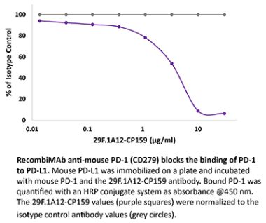
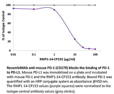
in vitroPD-1neutralization
Park,S.J.,et al.(2014)。“Negative role of inducible PD-1 on survival of activated dendritic cells”J Leukoc Biol95(4):621-629。PubMed
PD-1is a well-established negative regulator of T cell responses by inhibiting proliferation and cytokine production of T cells via interaction with its ligands,B7-H1(PD-L1)and B7-DC(PD-L2),expressed on non-T cells.Recently,PD-1was found to be expressed in innate cells,including activated DCs,and plays roles in suppressing production of inflammatory cytokines.In this study,we demonstrate that PD-1 KO DCs exhibited prolongevity compared with WT in the dLDCs of pads.Interestingly,uponLPS stimulation,WT DCs increased the expression of PD-1and started to undergo apoptosis.DCs,in spleen of LPS-injected PD-1KO mice,were more resistant to LPS-mediated apoptosisin vivothan WT controls.Moreover,treatment of blocking anti-PD-1mAb during DC maturation resulted in enhanced DC survival,suggesting that PD-1:PD-L interactions are involved in DC apoptosis.As a result,PD-1-deficient DCs augmented T cell responses in terms of igen-specifificient DCs augmented forations CD 4 and CD 8 T cells to a greaterdegree than WT DCs.Moreover,PD-1 KO DCs exhibited increased MAPK1and CD40-CD40L signaling,suggesting a possible mechanism for enhanced DC survival in the absence of PD-1 expression.Taken together,our findings further extend the function of PD-1,which plays an important roltope of des important implicationsfor PD-1-mediated immune regulation。
in vivoPD-1/PD-L信号
Cooper,Z.A.,et al.(2014)。“Response to BRAF inhibition in melanoma is enhanced when combined with immune checkpoint blockade”Cancer Immunol Res2(7):643-654。PubMed
BRAF-targeted therapy results in objective responses in the majority of patients;however,the responses are short lived(approximately6months)。In contrast,treatment with immune checkpoint inhibitors results in a lower response rate,but the responses tend to be more durable.BRaF inhibition results in a more favorable tumor microenvironment in patients,with an increase in CD8(+)T-cell infiltrate and a decrease in munoppressive cytokines。There is also increased expression of the immunomodulatory molecule PDL1,which may contribute to the resistance.On the basis of these findings,we hypothesized that BRAF-targeted therapy synergize with the PD 1pathway blockade to enhance antitumor immunity.To test this his hypothesis,wen Ptva(/-)syngeneic tumor graft immunocompetent mouse model in which BRaF inhibition leads to a significant increase in the intratumoral CD8(+)T-cell density and cytokine production,similar to the effects of BRaF inhibition in patients.In this model,CD8(+)T cells were found to play a critical role in the therapeutic effect of BRaF inhibition。Administration of anti-PD1 or anti-PDL1 together with a BRAF inhibitor led to an enhanced response,significantly prolonging survival and slowing tumor growth,as well as significantly increasing the number and activity of tumor-infiltrating lymphocytes.These results demonstrate synergbetweed联锁点,联锁点blockade.Although clinical trials combining these two strategies are ongoing,important questions still remain unanswered.Further studies using this new melanoma mouse model may provide therapeutic insights,including optimatal timing and sequence of therapy。
in vivoPD-1/PD-L信号,in vitroPD-1neutralization
Duraiswamy,J.,et al.(2013)。“Dual blockade of PD-1 and CTLA-4combined with tumor vaccine effectively restores T-cell rejection function in tumors”Cancer Res73(12):3591-3603。PubMed
Tumor progression is facilitated by regulatory T cells(Treg)and restricted by effector T cells。CD8(+)T cells and Foxp3(+)Tregs by programmed death-1(PD-1,PDCD1).In addition,we identify an additional role of CTL antigen-4(CTLA-4)inhibitory receptor in further promoting dysfunction of CD8(+)teffector cells in tumor models(CT26colon carcinoma and ID8-VEGF ovarian carcinoma)。Two thirds of CD8(+)tumor-infiltrating lymphocytes(TIL)expressed PD-1,whereas one third to half of CD8(+)TIL coexpressed PD-1and CTLA-4。PD-1(+)CTLA-4(+)CD8(+)TIL had characteristics of more severe dysfunction than single-positive(PD-1(+)or CTLA-4(+))TIL,including an inability to proliferate and secrete effector cytokines.Blockade of both PD-1and CTLA-4resulted in reversal of CD8(+)TIL dysfunction and led to tumor rejection in two thirds of mice。双封闭式配电盘CD8(+)和CD4(+)T cells,antigen-specific cytokine release,inhibition of suppressive functions of Tregs,and upregulation of key signaling molecules critical for T-cell function.When used in combination with GVAX vaccination(consisting of granulocyte macrophage colony-stimulating factor-expressing radiated turcells),inhibitory pathway blockade induced rejection of CT26tumors in100%of mice and ID8-VEGF tumors in75%of mice.Our study indicates that PD-1signaling in tumors is required for both suppressing effector T cells and maintaining tumor Tregs,and that PD-1/PD-L1 pathway(CD274)effector T-cell activity,thereby attenuating Treg suppression。
流动周期,流动周期
Good-Jacobson,K.L.,et al.(2012)。“CD80 expression on B cells regulates murine T follicular helper development,germinal center B cell survival,and plasma cell generation“J Immunol188(9):4217-4225。PubMed
Germinal center(GC)B cells and T follicular helper(T(FH))cells interact in the production of high-affinity long-lived plasma cells(PCs)and memory B cells,although the mechanisms regulating the formation of these long-lived populations remain unclear.Because CD 80is one of the few markers shared by human and murine memory B cells,we investigated its role in the development of GCs,memory cells,and PCs.In CD80-deficient mice,fewer long-lived PCwed commuzation thain B6controls.In concert,the absence of CD80resulted in an increase in apoptotic GC B cells during the contraction phase of the GC.CD80(-/-)mice had fewer T(FH)cells compared with that of B6,and residual T(FH)cells failed to mature,with decreased ICOS and PD-1expression and decreased synthesis of IL-21mRNA.Mixed bone marrow chimeras demonstrated a B cell-intrinsic requirement for CD80expression for normal T(FH)cell and PC development。Therefore,B cell expression of CD80plays a critical role in regulating B-T interactions in both early and late GC responses.This,in turn,results in impaired ability to produce long-lived PCs.These data provide new insights into the development of GCs and Ab-forming cells and the functions of CD80huin moration imnity。
Western Blot,Immunofluorescence
Chen,L.,et al.(2009)。“Role of the immune modulator programmed cell death-1 during development and apoptosis of mouse retinal ganglion cells”Invest Ophthalmol Vis Sci50(10):4941-4948。PubMed
PURPOSE:Mammalian programmed cell death(PD)-1is a membrane-associated receptor regulating the balance between T-cell activation,tolerance,and immunopathology;however,its role in neurons has not yet been defined.The hypothesis that PD-1signaling actively promotes retinal ganglion cell(RGC)death within the developing mouse retina was investigated。EthODS:Mature retinal cell types expressing PD-1 were identified by immunofluorescence staining of vertical retina sections;developmental expression was localized by immunostaining and quantified by Western blot analysis.PD-1 indevelopmental RGC survival was assessed edin vitrousing retinal explants andin vivousing PD-1knockout mice.PD-1ligand gene expression was detected by RT-PCR.RESULTS:PD-1is expressed in most adult RGCs and undergoes dynamic upregulation during the early postnatal window of retinal cell maturation and physiological programmed cell death(PCD)。In vitro blockade of PD-1signaling during this time selectively increases the survival of RGCs.Furthermore,PD-1-deficient mice show a selective increase in RGC number in the neonatal retina at the peak of developmental RGC death.Lastly,gene expression of the immune PD-1ligand gend nes Pdcds1出口出口出口总出口量调节器maturation.CONCLUSIONS:These findings collectively support a novel role for a PD-1-mediated signaling pathway in developmental PCD during postnatal RGC maturation。
The programmed death1/programmed death1ligand(PD-L)pathway is instrumental in peripheral tolerance。Blocking this pathway exacerbates experimental autoimmune diseases,but its role in autoimmune kidney disease has not been explored.Therefore,we tested the hypothesis that the programmed death1ligands(PD-L1and PD-L2),provide a protective barrier during T cell-and macrophage(Mphi)-dependent autoimmune kidney disease。Forthis purpose,we compared nephrotoxic serum nephritis(NSN)in mice lacking PD-L1(PD-L1(-/-),PD-L2(PD-L2(-/-)),or both(PD-L1/L2(-/-)to wild-type(WT)C57BL/6mice。Kidney pathology,loss of renal function,and intrarenal leukocyte infiltrates were increased in each PD-L(-/-)strain as compared with WT mice。Although the magnitude of renal pathology was similar in PD-L1(-/-)and PD-L2(-/-)mice,our findings suggest that kidney disease in each strain is regulated by distinct mechanism.Specifically,we detected increased CD68(+)cells along with elevated circulating IgG and IgG deposits in glomeruli in PD-L2(-/-)mice,but not PD-L1(-/-)mice。In contrast,we detected arise in activated CD8(+)T cells in PD-L1(-/-)mice,but not PD-L2(-/-)mice。Furthermore,since PD-L1is expressed by parenchymal and hemopoietic cells in WT kidneys,we explored the differential impact of PD-L1expression these cell types by inducing NSN in bone marrow chimeric mice.Our results indicate that PD-L1expression on hemopoietic cells,and not parchenricells,priss ble for limiting leukocyte infiltration during NSN.Taken together,our findings indicate that PD-L1and PD-L2provide distinct negative regulatory checkpoints poised to suppress autoimmune renal disease。
in vivoPD-1/PD-L信号
Barber,D.L.,et al.(2006)。“Restoring function in exhausted CD8T cells during chronic viral infection”Nature439(7077):682-687。PubMed
Functional impairment of antigen-specific T cells is a defining characteristic of many chronic infections,but the underlying mechanisms of T-cell dysfunction are not well understood.To address this question,we analysed genes expressed in functionally impaired virus-specific CD8 T cells refects in inhompy cytic choriomeningitis virus(LCMV),and compared these with the gene profile of functional memory CD8T cells.Here we report that PD-1(programmed death1;also known as Pdcd1)was selectively upregulated by the exhausted T cells,and thatin vivoadministration of antibodies that blocked the interaction of this inhibitory receptor with its ligand,PD-L1(also known as B7-H1),enhanced T-cell responses.Notably,we found that even in persistently infected mice that were lacking CD4T-cell help,blockade of the PD-1/PD-L1 inhibitory pathway had a beneficial effection on the‘helpless’CD8T cells,restoring their ability to undergo proliferation,secreteed and ected se viral load.Blockade of the CTLA-4(cytotoxic T-lymphocyte-associated protein4)inhibitory pathway hadno effect on either T-cell function or viral control。These studies identify a specific mechanism of T-cell exhaustion and define a potentially effective immunological strategy for the treatment of chronic viral infections。
Immunohistochemistry(frozen)
Liang,S.C.,et al.(2003)。PD-1,PD-L1,and PD-L2expression during normal and autoimmune responses“Eur J Immunol33(10):2706-2716。PubMed
Newer members of the B7-CD28 superfamily include the receptor PD-1and its two ligands,PD-L1and PD-L2.Here,we characterize the expression of PD-1,PD-L1,and PD-L2 in tissues of naive miceand in target organs from two models of autoimmunity,the pancreas fromnon-obesand intarget organs from地下排水量alomyelitis(EAE)。In naive mice,proteiexpression of PD-1,PD-L1,and PD-L2was detected in the thymus,while PD-1and PD-L1were detected in the spleen.PD-L1,but not PD-L2,was also detected at low levels on cardiac endothelium,pancreatic islets,and syncyciotrophoblastin the notic mice,PD-1 and PD-L1 were expressed on infiltrating cells in the pancreatic islets.Furthermore,PD-L1was markedly up-regulated on islet cells.In brains from mice with EAE,PD-1,PD-L1,and PD-L2were expressed on infiltrating inflammatory cells,and PD-L1was up-regulated on endothelium within EAbrain.Thedistression of PD-L2 led us to compare their transcriptional reSTAT4(-/-),STAT6(-/-),orNF-kappaB p50(-/-)p65(+/-)dendritic cells(DC)。PD-L2,but not PD-L1,expression was dramatically reduced in p50(-/-)p65(+/-)DC。Thus,PD-L1and PD-L2exhibit distinct expression patterns and are differentially regulated on the transcriptional level。